
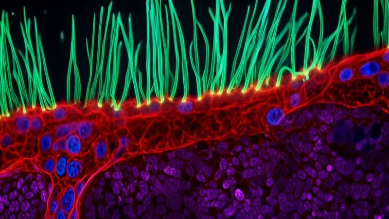
Microscopic Video of Lung Cilia Wins 2026 Nikon Small World in Motion Prize
Sandego.net – A close-up view of tiny structures inside a child’s airways has earned the top honor in the 16th annual Nikon Small World in Motion competition, turning a difficult respiratory condition into a striking moving image.
The winning footage was created by Ning Xu of Tsinghua University in Beijing, China. Using advanced microscopy, Xu recorded the unusual movement of airway cilia in a child with primary ciliary dyskinesia, also known as PCD. The competition recognizes videos that reveal subjects beyond the range of normal human vision, combining scientific observation with visual impact.
Why the movement matters
Cilia are extremely small, hairlike projections found on many cells in the body. In the airways, they normally move in coordinated patterns that help transport mucus and trapped debris out of the lungs. Their motion is an important part of the body’s natural airway-cleaning system.
For people with primary ciliary dyskinesia, this action can be disrupted. Rather than moving in an effective, coordinated rhythm, the cilia may beat abnormally. That reduced ability to clear material from the airways can contribute to repeated respiratory infections.
Xu’s video makes that difference visible through motion rather than a single static frame. The footage captures the intricate activity of cilia and illustrates why moving microscopy can be especially valuable for examining conditions involving cellular behavior.
“To understand this disease, you need to observe around 10 seconds of motion, not just a still image,” Xu, an optical engineer, said in a press release Wednesday.
Xu develops imaging systems intended to help researchers study complex cell structures through movement. The winning entry highlights how a short sequence can reveal patterns that would be difficult or impossible to recognize in a photograph alone.
“When people watch cilia beating, they immediately understand that something beautiful and important is happening inside us,” Xu added. “I think this competition turns microscopy into a shared language.”
A competition built around the unseen
Nikon Small World in Motion was established to celebrate advances in microscopic photography and video. The contest is open to people interested in photography or video, and its entries span scientific subjects that are ordinarily too small to be seen without specialized equipment.
This year’s video competition attracted 347 submissions from 40 countries. A judging panel made up of experts in science and advanced light microscopy selected the winners.
Microscopy can serve several purposes at once: it can help researchers examine living systems, document processes over time and make scientific ideas more understandable to wider audiences. Videos such as Xu’s offer viewers a direct look at activity happening within the human body at a scale far removed from everyday experience.
The visual appeal of microscopic imaging is also central to the contest. Minute structures, cells and organisms can appear highly abstract when magnified, even though they are part of familiar biological processes. In this case, the motion of airway cilia provides both a compelling image and a clear illustration of why their function matters for lung health.
“Xu’s work, and the work of all our winners, reflects the competition’s enduring purpose to inspire wonder, fuel discovery, and showcase the artistry inherent in scientific exploration,” said Eric Flem, senior manager of communications and CRM at Nikon Instruments.
Other winning microscopic scenes
The remaining honorees captured a broad range of biological activity. Second place went to Nguyen Nam Nhat for a video featuring a tiny roundworm alongside a single-celled Dileptus organism.
Benedikt Pleyer received third place for footage of jellyfish larvae suspended in water droplets. The video presents an aquatic subject in a setting where the droplets themselves become part of the visual composition.
Fourth place was awarded to Andrew Moore, whose entry showed neighboring cells dividing in synchronization. Cell division is fundamental to growth and repair, and the video focuses on the coordinated timing of that process across adjacent cells.
Patrick Hickey took fifth place with an image sequence from turtle vine leaf cells. His footage showed mitochondria in green and chloroplasts in red, emphasizing the movement and changing arrangement of structures within plant cells.
Motion brings hidden biology into view
The 2026 winners demonstrate the range of stories that can unfold under a microscope: respiratory cells moving within an airway, organisms interacting at a tiny scale, developing jellyfish larvae, dividing cells and active structures inside plant leaves.
For Xu’s winning work, the central subject is not simply the appearance of cilia but the way they move. That distinction is significant because cilia are designed to work collectively. Their usual coordinated beating helps maintain clearer airways, while abnormal motion can interfere with that role in people with PCD.
By capturing that activity over several seconds, the video gives viewers an accessible way to see a process that is normally hidden. It also reinforces the value of scientific imaging as a bridge between specialized research and public understanding, showing that some of the body’s most consequential activity takes place at a scale the eye cannot reach.
Related Reading
Frequently Asked Questions
What is Astonishing footage of abnormal cell movement?
Astonishing footage of abnormal cell movement is the main topic of this guide. The article explains the context, practical details, and next steps readers should understand.
Why does Astonishing footage of abnormal cell movement matter?
Astonishing footage of abnormal cell movement matters because readers are looking for a useful answer, not just a short summary. Good content should match search intent and help them decide what to do next.